Cellophane banding vs ameroid ring constrictors for portosystemic shunts
What you need to know about these two common methods of portosystemic shunt attenuation in dogs and cats.
Cellophane versus ameroid ring constrictors: two sides of the same coin.
Two of the most common methods of slowly attenuating congenital portosystemic shunts in dogs and cats are cellophane banding and ameroid ring constrictors. These modalities are similar in that they both aim to slowly cause attenuation of the portosystemic shunt vessel in order to avoid complications such as portal hypertension and development of medusa-like acquired shunting.
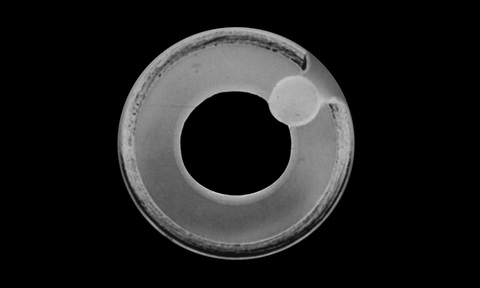
Ameroid ring constrictors are made up of an outer rim of stainless steel, which encases a 'C' shaped doughnut of casein, which is a highly hygroscopic milk protein derivative. This casein achieves two things which makes it appropriate for slowly attenutating portosystemic shunts.
Firstly, casein absorbs bodily fluids within the peritoneal cavity which forces it to expand, however the outer metallic rim forces the expansion inward. A study by Hunt et al (Veterinary Surgery, 2014) found that by 9 weeks post implantation, the ameroid ring constrictor only closed to 42-48% of the original internal diameter. Whilst this seems to be a mediocre outcome, clinically nearly all dogs do well, as the constrictor causes thrombosis of the shunting vessel. Hunt's study also suggested that ameroid rings >5mm internal diameter may be at slightly more risk of allowing continued shunting.
The second means by which ameroid rings cause attenuation of the portosystemic shunt is by a perivascular foreign body reaction induced by the casein. Based on Hunt's 2014 study, the fibrosis caused by the casein may be as equally important as physical extraluminal constriction around the shunting vessel. It is likely that both the compression and perivascular fibrosis mechanisms dovetail to induce tamponade and thrombosis of the shunting vessel to achieve clinical results.
Ameroid rings, whilst effective in eliminating clinical signs in 92% of extraheptic portosystemic shunt cases (Falls, Veterinary Surgery, 2013), are reported to allow continued shunting in 18-24% of cases in which they are applied (Falls, 2013 and Hunt, 2014). Long term outcomes in cases that have ameroid rings placed however are still excellent, with median survival times of 12 years (Falls, Veterinary Surgery, 2013).
Despite the risk of persistent shunting due to partial closure, ameroid are excellent for most dog and cat patients with portosystemic shunts. In certain instances however, alternative methods of attenuation such as cellophane banding are required. As we have discussed earlier, real cellophane is made from extruded regenerated cellulose. Before CelloVet, sourcing cellophane for use in veterinary surgery was haphazard. Veterinary surgeons were known to buy cigarettes, flowers or greeting cards in the hopes that the packaging they were supplied in was real cellophane. As Smith (Veterinary Surgery, 2014) has shown, it is very unlikely that the material peddled as cellophane in your local store is cellulose based, but more likely a plastic. Moreover, even if you stumble across what may be a cellulose based film, 99.9% of the time it will have some sort of plastic or adhesive coating to improve its material properties, handling and shelf life. At CelloVet we provide pure, uncoated, cellulose based cellophane which is free from any additional treatments.
So how does cellophane clinically perform in veterinary patients with congenital portosystemic shunts? One of the first descriptions of technique was by Drs. Youmans and Hunt (AVJ, 1998) in eleven dogs. Clinical outcomes in all dogs was excellent with resolution of signs, and 10/11 had sonographic evidence of complete attenuation of the shunt. In 2004, Dr. Hunt et al reported the clinical outcomes of 106 dogs and 5 cats that had cellophane banding for portosystemic shunting. Post-operative survival was excellent, with all cats and 94.5% of dogs being discharged from hospital. There were increased complications in the dogs which had intrahepatic shunts vs extraheptic, however if the animal survived to discharge, clinical signs resolved in all patients. Furthermore, of the available post-op 88 cases which had ammonia tolerance tests, 84% were normal and 16% were abnormal irrespective of the fact that all were clinically normal.
Debate remains as to how to best apply cellophane around both extra-heptic and intra-hepatic shunts. Frankel et al (JAVMA. 2006) evaluated outcomes in dogs that had cellophane banding placed around their shunts with and without attenuation of the vessel by the band to <3 mm. Attenuating the shunt to <3 mm was based upon recommendations first made by Youmans and Hunt (AVJ, 1998), however the study by Frankel et al (JAVMA, 2006) questions whether this arbitrary diameter of shunt attenuation is required. This study found no difference in clinical outcomes, however by 6 months post-op, post-prandial serum bile acid assays were within normal limits in the group which only had the cellophane gently wrapped around the vessel rather than securing the cellophane in place whilst attenuating the vessels to <3 mm. It should be noted however that most dogs in the group that did have cellophane attenuate the vessel to < 3 mm at the time of surgery likely had complete attenuation by 2.25 months, however this group also had increased serum bile acids at 6 months which suggests that the risk of acquired shunt development may be increased in animals which have the vessel attenuated arbitrarily to <3 mm at the time of surgery. Similar findings were made by Landon et al (Australian Veterinary Journal, 2008) in 16 dogs which had cellophane attenuation to <3 mm. In that study transcolonic scintigraphy post-op was used to identify continued shunting in 3/16 (18.6%) and multiple acquired shunts in 3/16 (18.6%) of cases. These results, along with Frankel et al (JAVMA, 2006) suggest that perhaps attenuation of the shunt to <3 mm may not be required as the risk for continued or acquired shunting is moderate. It stands to reasons that if attenuation of the shunting vessel is required intra-op, then portal pressures should be measured to help guide the degree of attenuation to reduce the risk of portal hypertension and acquired shunting.
Cellophane banding in cats has been reported by both Hunt et al (2004, Veterinary Surgery) and Cabassu et al (2011, JAVMA). In the report by Cabassu, 9 cats had cellophane banding for portosystemic shunts. There were 3 cats which died or were euthanized in the peri-operative period from neurological complications. Of the surviving cats, 5 had serum bile acids within normal limits and 4 were clinically normal by 3 months post-op. Anecdotal claims that cats have a poorer outcome compared to dogs when their shunts are attenuated using cellophane banding are unfounded. Cellophane has been used successfully in cats and dogs alike, and no difference in cat's ability to form a mounted foreign body reaction around cellophane within the peritoneal cavity has been proved.
Thankfully for the surgeon, the most mechanically secure means of cellophane banding has been evaluated by McAlinden et al (Veterinary Surgery, 2010). This study found that a maximum of 4 large (relative to the width of the cellophane strip) metallic vascular clips applied in alternating directions was the most secure. Furthermore, this study recommended that the cellophane be folded into at least 3 layers to help improve mechanical stability of the clips.
Whether you choose an ameroid ring or cellophane for your patient, first consider the following in order to aid your decision making.
For these reasons it is important to understand the benefits and limitations of both ameroids and cellophane for portosystemic shunt attenuation in dogs and cats. CelloVet was founded with the idea that surgeons should be able to have a choice of implant and be certain that the product they are using is pure, unadulterated cellulose derived cellophane. Be certain it is cellophane - use CelloVet.
Also in CelloVet | Cellophane for portosystemic shunts
Follow
© 2026 CelloVet.
Pty Ltd. All rights reserved. CelloVet is a trademark of CelloVet Pty. Ltd. PO Box 726, Fremantle, Western Australia 6959. Distributed by TPLOnly Inc., Canada.
Powered by Shopify